
If you've been treating dark spots as sun damage but nothing seems to fade them, you might be aiming at the wrong target. Not all hyperpigmentation is simply sun-related. A specific type—often called post-inflammatory hyperpigmentation—is triggered by inflammation in the skin rather than UV exposure alone. Understanding the difference changes how you approach treatment.
Dermatologists explain that when skin cells become inflamed, they produce excess melanin in a targeted area. The result looks similar to sun spots, but the cause is internal skin distress, not cumulative UV rays. Recognizing the warning signs early helps you stop the cycle before those marks set in deep.
1. The spots appeared after a pimple, rash, or irritation
Think back to the first time you noticed the discoloration. Did it follow a breakout, an allergic reaction, or even an aggressive facial? If you can trace the dark mark directly back to a blemish, scratch, or inflammation event, that's a strong indicator your hyperpigmentation is post-inflammatory, not photo-induced.
Sun spots develop gradually over years of exposure and tend to appear on areas that get the most direct sunlight—like the cheeks, nose, and shoulders. In contrast, post-inflammatory hyperpigmentation shows up exactly where the inflammation happened, even in places that rarely see the sun.
2. The edges are fuzzy, not sharply defined
A sun spot tends to have a clear, rounded border. It's often a flat, well-circumscribed patch of tan or brown. Post-inflammatory hyperpigmentation, however, often looks softer at the edges. The area may seem to feather out into the surrounding skin, and the color can shift from light brown to reddish-purple, especially if the inflammation is still active.
If your skin has a bit of residual redness or a dusky tone around the dark mark, that's another clue. Active inflammation leaves a trail. Sun damage alone doesn't carry that pink or purple undertone once the tan has settled.
3. The spots are on areas where you get breakouts or friction
Post-inflammatory pigmentation loves repetitive skin stress. If you consistently see dark patches along your jawline where acne flares, on your temples where your glasses rest, or on areas where you shave or wax, the root cause is likely inflammation, not the sun. Friction, picking at skin, and even strong chemical peels can trigger this response in melanin-rich skin.
People with darker skin tones—Fitzpatrick types IV through VI—are particularly prone to this type of pigmentation because their melanocytes are more reactive to any kind of inflammatory trigger. For them, a simple scratch or a mild heat rash can leave a lingering dark mark that lasts for months.
Why treatment differs for inflammatory vs. sun pigmentation
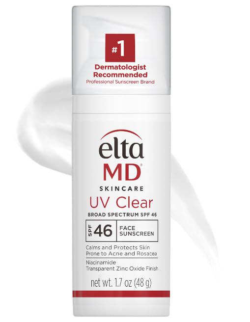
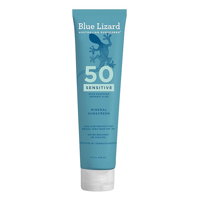
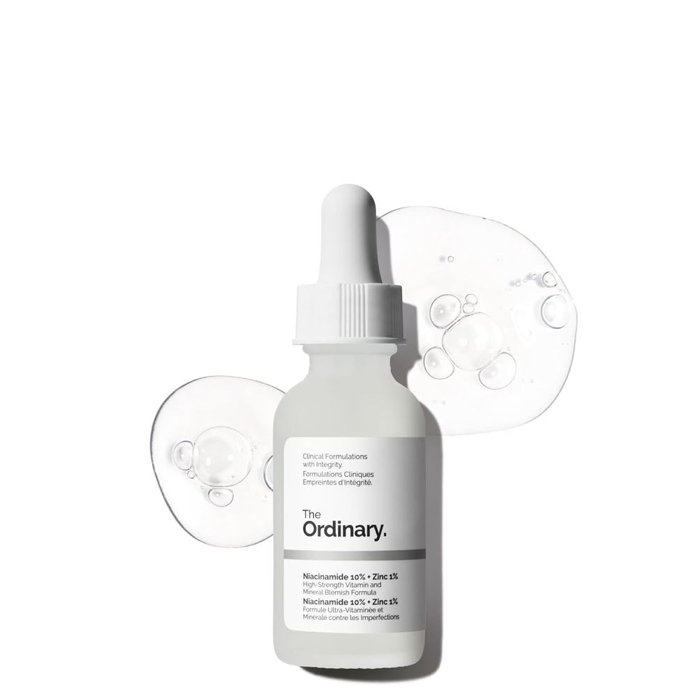
Treating post-inflammatory hyperpigmentation starts with stopping the inflammation first. Sunscreen still matters—UV rays can darken any existing spot—but the primary strategy is calming the skin. Products with azelaic acid, niacinamide, tranexamic acid, and gentle antioxidants tend to work better than aggressive acids or laser treatments, which can worsen inflammation in reactive skin.
Sun-related spots, on the other hand, respond well to tyrosinase inhibitors like kojic acid, vitamin C, and retinoids paired with strict sun avoidance. If you apply a formula designed for sun spots to an inflammatory spot, you may actually irritate the area and deepen the discoloration.
A simple rule: if the area still feels slightly warm, tender, or itchy, treat the inflammation before you treat the pigment. Once the skin is calm, the melanin will fade more predictably.
How to confirm your skin's issue at home
Take a photo of the spot in natural light and examine the color. Sun spots typically look uniform tan or brown. Inflammatory pigmentation often has a reddish, purplish, or grayish hue alongside the brown. If you press gently on the spot and it blanches slightly, there's likely still active inflammation beneath the surface.
Also note the timeline. Sun spots appear slowly and stay. Inflammatory marks tend to fade faster once the trigger is removed—usually within three to six months if you stop picking at acne or change your skincare routine. If the dark spot has not changed shape or color after six months of gentle care, a dermatologist can assess it with a handheld dermoscope to rule out melanoma or other growths.
Knowing whether your hyperpigmentation is driven by sun or inflammation changes your entire skincare strategy. Treating the wrong type can waste months of effort and even darken the area. Pay attention to how the spot appeared, what it looks like up close, and whether your skin is still reactive around it. Those three clues point you toward the right approach—and real progress.