
You wash your face, you wear sunscreen on sunny days, and you avoid tanning beds. Yet one morning, there it is: a flat, brown spot on the back of your hand that wasn't there last year. Age spots, sometimes called liver spots or solar lentigines, have a way of sneaking up on us. But the name itself is misleading. The real root cause is not how many birthdays you've had. To understand what actually drives these spots, it helps to look at what dermatologists see under the microscope and in the clinic every day.
What are age spots, exactly?
An age spot is a localized area of increased pigment. It happens when melanocytes—the pigment-producing cells in your skin—start to produce too much melanin in one spot. Unlike a freckle, which fades when you avoid the sun, an age spot is stubborn. It builds up over years because the skin cells that hold that pigment, called keratinocytes, keep accumulating melanin granules. The spot itself is flat, usually round or oval, and ranges from light tan to dark brown. It is completely benign, but it tells a story about cumulative exposure.
The real culprit: ultraviolet exposure
Dermatologists consistently point to ultraviolet radiation as the primary driver. It's not aging itself that creates the spot; it's the sum of every moment your skin was exposed to UV rays without protection. A 30-minute walk at noon ten years ago, a long beach vacation in your twenties, or even the reflected UV from a snowy mountain—each event adds to a cellular memory of damage. The skin reacts by trying to shield itself. It sends extra melanin to the surface to absorb and scatter UV rays. Over decades, that defense mechanism becomes uneven. Some areas produce too much pigment, and the body's ability to regulate melanin production gradually weakens.
Think of UV exposure as a photocopier that makes extra copies every time you press start. After hundreds of small exposures, the page gets messy.
Why the face and hands show the most spots
Look at where age spots commonly appear: the backs of hands, forearms, face, shoulders, and upper chest. These are the areas that get the most sun over a lifetime. The back of a hand, for instance, is exposed almost every time you drive, hold a coffee cup, or walk outside—even if you don't think of it as a sun-exposed spot. The same goes for the sides of the forehead or the tops of the ears. Dermatologists use this pattern to differentiate age spots from other pigmentation issues like melasma or post-inflammatory hyperpigmentation, which follow different patterns.
Lifelong cumulative exposure vs. aging itself
There is a common misconception that skin simply ages and spots appear automatically. In controlled studies, people who protect their skin from the sun from an early age show significantly fewer age spots, even in their seventies. In contrast, individuals with high lifetime sun exposure often develop these spots in their thirties or forties. That wide age range is a clue. The trigger is not chronological age; it's the total amount of UV energy that has hit the skin. Genetics does play a role—some people have melanocytes that are more reactive to UV—but even for them, sun avoidance reduces the result.
Artificial UV sources also contribute. Tanning beds deliver concentrated UVA and UVB radiation. UVA, in particular, penetrates deeply into the dermis and triggers long-term changes in melanocyte activity. One study published in the Journal of the American Academy of Dermatology found that women who used tanning beds had a significantly higher number of age spots on their arms and trunk compared to non-users, regardless of their age.
Hormones and inflammation: the secondary triggers
While UV is the central actor, it doesn't act alone. Hormonal shifts—especially during pregnancy or while taking oral contraceptives—can increase melanin production throughout the body. This can make existing age spots appear darker, or new ones may emerge on the face. This condition, often called melasma, is a different beast from age spots, but the two can overlap. Inflammation from a cut, a pimple, or an allergic reaction can also leave a dark mark that resembles an age spot. Known as post-inflammatory hyperpigmentation, this is more common in darker skin tones and can be confused with lentigines if not examined closely. However, true age spots (solar lentigines) are specifically linked to UV-driven hyperplasia of melanocytes, not transient inflammation.
How dermatologists distinguish an age spot from something else
During a skin exam, a dermatologist uses a tool called a dermatoscope—a magnifying lens with a light—to look at the spot's pigment network. An age spot shows a uniform, delicate honeycomb or reticular pattern under the scope. A mole or an early melanoma, in contrast, often shows irregular dots, globules, or chaotic pigment. If there is any asymmetry, irregular borders, more than one color, or a diameter larger than a pencil eraser, a biopsy might be done. It is worth remembering that even common spots that change shape or color need professional evaluation.
The golden rule: any spot that looks different from your other spots—or that changes, itches, or bleeds—deserves a check. Trust your eyes, and see a board-certified dermatologist.
What about oxidative stress and internal factors?
Newer research points to oxidative stress as an amplifier. UV radiation creates free radicals in the skin, which damage DNA and trigger signaling pathways that stimulate melanin production. Antioxidants in the skin (like vitamin C and E) can neutralize some of these free radicals, but prolonged UV exposure depletes them over time. That is why topical antioxidants are often recommended as a second line of defense after sunscreen. However, taking high-dose antioxidant supplements has not been proven to reverse existing age spots. Internal factors like diabetes or thyroid disorders can sometimes cause hyperpigmentation, but those conditions produce a diffuse darkening, not the discrete, well-defined spots of solar lentigines.
Can age spots be prevented or minimized?
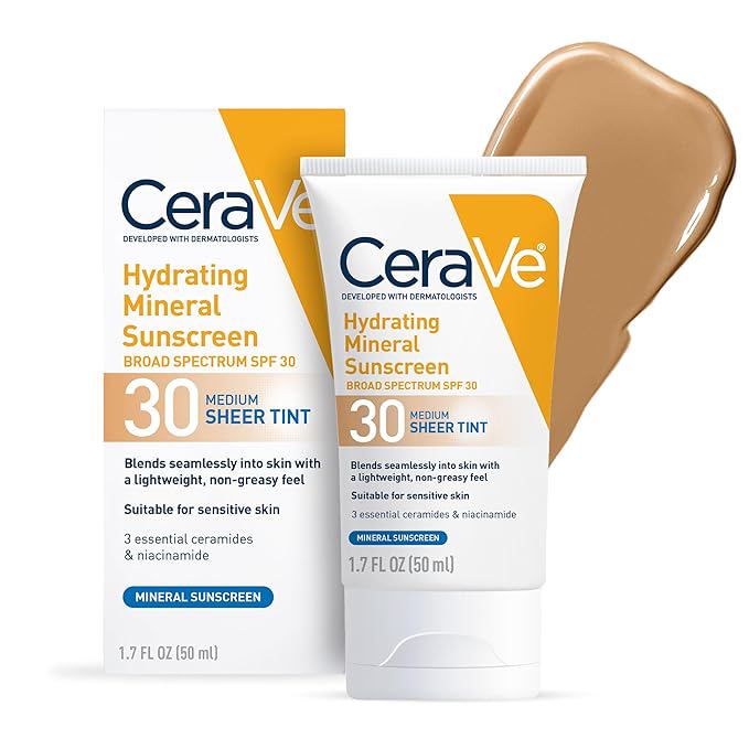
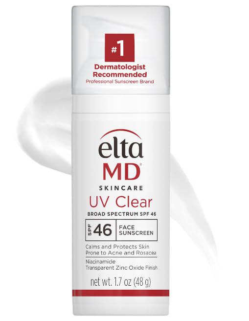
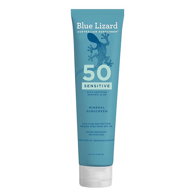
Prevention centers on minimizing cumulative UV exposure. A broad-spectrum sunscreen with SPF 30 or higher, applied to all exposed skin daily, is the first step. However, one small nuance: many people skip the backs of their hands when applying sunscreen. A pea-sized dab on the face and nothing on the hands means the hands accumulate years of unprotected exposure. Making hand sunscreen a habit—reapplying after hand washing—is a simple change that can affect outcome over time. Beyond sunscreen, sun-protective clothing, wide-brimmed hats, UV-blocking sunglasses, and seeking shade during peak hours from 10 AM to 4 PM all contribute to reducing total UV burden.
If age spots are already present, options like topical retinoids, hydroquinone, or chemical peels may lighten them under a dermatologist's care. Laser treatments, including Q-switched or picosecond lasers, are effective at targeting the pigment without damaging the skin's surface. A specific laser called the KTP or the IPL (intense pulsed light) can also reduce contrast. But these treatments work best when the underlying habit of sun protection is firmly in place. Without it, the spots will return within months.
One simple takeaway that dermatologists stress: age spots are a sign of past sun exposure, not a verdict of aging. The body's ability to protect and repair itself remains strong at any age. With daily sun protection and the right clinical care, skin can maintain a more even tone even after decades in the sun.