
Learning that you or your child may have a congenital heart defect (CHD) can feel overwhelming. The path from suspicion to diagnosis is often paved with many tests and specialist visits, but understanding what each step involves can help you feel more prepared and less anxious. This guide walks through the standard diagnostic journey, from the first prenatal ultrasound through the later stages of life, focusing on what patients and families can expect.
A congenital heart defect is a structural problem with the heart that is present at birth. These defects range from simple holes between chambers to complex malformations of valves or major blood vessels. While some are detected during pregnancy, others may not appear until childhood or even adulthood. The diagnostic process is designed to be as thorough as it is careful, using a combination of physical exams, imaging, and monitoring.
Prenatal screening: the first look
For many families, the first hint of a CHD comes during a routine pregnancy ultrasound. Around the 18th to 22nd week of gestation, a detailed anatomy scan examines the baby's heart structure. The sonographer looks for four distinct chambers and checks that major blood vessels connect properly. If something appears unusual, the next step is typically a fetal echocardiogram.
A fetal echo is a specialized ultrasound that provides a much closer look at the heart's anatomy and blood flow. It is non-invasive and carries no risk to the mother or baby. The results can confirm a defect, rule one out, or simply suggest that further monitoring is needed. Some defects, however, are not visible in the womb because they develop or become apparent only after birth when circulation patterns change.
Newborn and infant diagnosis: listening and looking
After birth, the first sign of a heart problem is often a heart murmur—a whooshing or swishing sound heard through a stethoscope. While many murmurs are innocent, some point to a structural issue. Your pediatrician or cardiologist will pay close attention to other clues too: rapid breathing, poor feeding, bluish tint to the skin or lips (cyanosis), or failure to gain weight.
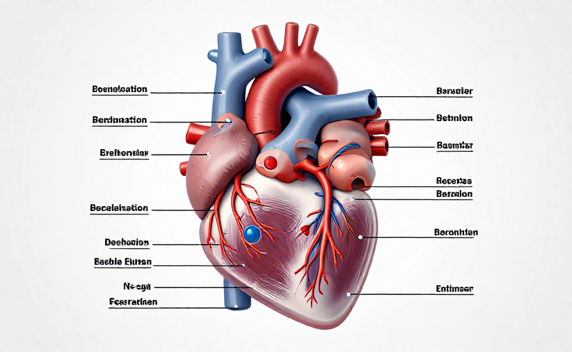
If a defect is suspected, the standard initial test is an echocardiogram, or echo. This ultrasound of the heart shows moving images of the heart valves, chambers, and pumping action. It is painless and usually takes about 45 minutes to an hour. The echo is the cornerstone of CHD diagnosis because it can reveal the exact location and size of most structural abnormalities.
An echocardiogram is the single most important tool for diagnosing congenital heart defects. It provides real-time, detailed images without any radiation exposure.
Additional diagnostic tools
Depending on what the echo shows, your cardiologist may recommend other tests to get a clearer picture. An electrocardiogram (ECG or EKG) records the heart's electrical activity and can identify rhythm problems or signs of enlargement. It is quick and involves placing small stickers on the chest. A chest X-ray can show the size and shape of the heart and whether there is extra fluid in the lungs, which can indicate heart failure.
For more complex defects, a cardiac MRI may be used. This is a detailed imaging scan that uses a magnetic field and radio waves to create 3D images of the heart. It is especially helpful for looking at blood vessels and the function of the right ventricle. Another key test is a Holter monitor, a portable device worn for 24 to 48 hours that continuously records the heart rhythm to catch intermittent problems.
In some cases, a cardiac catheterization is necessary. This is a minimally invasive procedure where a thin tube is threaded through a blood vessel in the leg or arm up to the heart. It measures pressures and oxygen levels inside the heart chambers and can also see blockages or abnormal connections. Sometimes, a minor intervention like closing a small hole can be done during the same procedure.
Diagnosis in older children and adults
Not all congenital heart defects are found early. Some mild defects, like a small atrial septal defect (ASD) or a bicuspid aortic valve, may not cause noticeable symptoms until later in life. Adults may first learn about a CHD after experiencing shortness of breath during exercise, palpitations, or an abnormal heart sound during a routine physical. The diagnostic process is essentially the same as in children, starting with an echo and building from there.
For adults with a known CHD who had childhood surgery, lifelong monitoring is key. Even after a successful repair, the anatomy is not normal, and issues like valve leakage, arrhythmias, or conduit narrowing can develop over time. Regular follow-up with a cardiologist who specializes in adult congenital heart disease is essential.
What the results mean
Diagnoses from these tests fall into categories based on complexity. Simple defects might include small holes like a ventricular septal defect (VSD) or a mild valve problem. Moderate defects could involve more significant structural changes, such as tetralogy of Fallot or coarctation of the aorta. Complex defects include single-ventricle conditions like hypoplastic left heart syndrome (HLHS) or transposition of the great arteries.
Your care team will explain which specific defect you or your child has, how it affects circulation, and the typical prognosis. No two cases are exactly alike, and treatment plans are highly individualized, ranging from monitoring alone to medications, catheter-based closure, or open-heart surgery.
What to ask your doctor
Being prepared for appointments can help you feel more in control. Consider asking these questions: What is the exact name of the defect? How will it affect day-to-day life and physical activity? What monitoring or follow-up is needed? Are there any activity restrictions? For a child, ask about growth, feeding challenges, and school sports. For yourself as an adult, ask about pregnancy risks and how the condition may change with age.
Every diagnostic test has a purpose, and each result brings you closer to a clear picture. The goal is not just to name the defect, but to create a roadmap for care that supports the best possible quality of life. Trust your medical team, take notes, and never hesitate to ask for a second opinion if something does not feel right. You are your own or your child's best advocate.